

The microcirculation, consisting of the smallest blood vessels within the body, plays a vital role in delivering oxygen and nutrients to tissues while removing metabolic waste products. Changes in microvascular blood flow can provide valuable insights into tissue health, vascular function, wound healing, inflammation, and disease progression. As a result, accurate assessment of blood flow has become increasingly important in both clinical research and healthcare applications.
Traditional methods of assessing tissue perfusion often provide limited information or require direct contact with the tissue. In contrast, modern imaging technologies allow clinicians and researchers to visualise blood flow across larger tissue areas, helping them better understand physiological responses and identify abnormalities that may not be apparent through conventional assessments alone.
How Laser Speckle Contrast Imaging Works

Laser Speckle Contrast Imaging (LSCI) is a non-contact imaging technique used to assess tissue blood flow in real time. The technology works by illuminating tissue with coherent laser light, creating a speckle pattern on the camera sensor. When blood cells move within the tissue, the speckle pattern changes. Faster blood flow results in greater blurring of the speckle pattern, while slower blood flow produces sharper images. By analysing these changes, the system generates colour-coded maps that represent tissue perfusion.
One of the key advantages of LSCI is its ability to capture full-field blood flow images at high speed while maintaining excellent spatial resolution. This allows users to observe dynamic changes in perfusion across an entire area of interest rather than relying on single-point measurements. As a result, LSCI provides a more comprehensive view of microvascular function and tissue viability.
Applications of LSCI in Research and Clinical Practice
The ability to visualise and quantify blood flow in real time has made LSCI a valuable tool across a wide range of research and clinical fields. In neuroscience, it is used to study cerebral blood flow and stroke models. In dermatology, it supports investigations into skin inflammation, irritancy, and vascular responses. Researchers also utilise LSCI in oncology, pharmacology, cardiovascular studies, and plastic surgery to better understand tissue perfusion and physiological changes.
Because LSCI is a non-contact technique, it is particularly useful when assessing sensitive tissues or situations where maintaining sterility is important. The technology can also be used to monitor responses to interventions such as pressure cuff testing, skin heating protocols, and pharmacological challenges. Its ability to provide objective, reproducible measurements makes it a valuable tool for both research and translational clinical applications.
Advancing Microvascular Assessment with the Moor FLPI-2
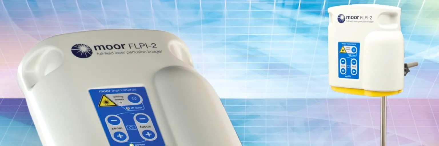
The Moor FLPI-2 is a Laser Speckle Contrast Imaging system designed to provide real-time, high-resolution assessment of tissue blood flow. Using advanced laser speckle contrast analysis, the system captures full-field perfusion images at video frame rates, allowing users to visualise dynamic changes in blood flow with exceptional clarity.
Featuring a non-contact design, motorised optical zoom, auto-focus capabilities, and flexible imaging areas, the FLPI-2 is suitable for a broad range of research and clinical applications. The system also includes powerful analysis software that enables region-of-interest assessment, data visualisation, reporting, and export functions. By combining speed, precision, and ease of use, the Moor FLPI-2 helps researchers and clinicians gain deeper insights into microvascular function and tissue perfusion.
More Information
- Moor Instruments. Laser Speckle Contrast Imaging (LSCI) – moorFLPI-2 Product Page. https://www.moor.co.uk/products/imaging/laser-speckle-contrast-imager/
Moor Instruments, BMEC Pte Ltd